Enhancing Anesthesia Practice with Immersive Technology: Anatomy, XR Imaging and Virtual Collaboration
Immersive Technology in the Field of Anesthesia
Anesthesiologists can leverage immersive technologies to enhance their understanding of regional anesthesia, sonoanatomy, and pre-surgical planning. By utilizing immersive 3D anatomy models, they gain a deeper insight into anatomical structures, which significantly improves their ability to identify nerve pathways, vascular structures, and fascial planes that are crucial for ultrasound-guided procedures. Moreover, this advanced visualization allows them to virtually dissect the body layer by layer, offering a comprehensive understanding of the anatomical planes—axial, sagittal, and coronal.
Enhancing Collaboration and Decision-Making
In addition to individual learning, immersive technology fosters real-time collaboration in a shared virtual space. This feature allows anesthesiologists to discuss cases, refine techniques, and plan procedures remotely, effectively bridging geographical gaps and enhancing teamwork. Furthermore, the integration of 3D Organon XR Imaging plays a pivotal role in improving decision-making. For example, by visualizing patient-specific CT and MRI scans in 3D before surgery, specialists are able to refine their approach, leading to greater precision in procedures and better overall patient outcomes.
A New View on Dissection with 3D Organon
3D Organon Anatomy provides anesthesiologists with an advanced, interactive tool that enhances their understanding of human anatomy and improves clinical decision-making. The module offers access to detailed anatomical structures across various body systems, enabling users to explore the complex relationships between tissues, organs, and vessels. These features are essential for anesthesia practice, as they allow anesthesiologists to visualize and analyze anatomical intricacies that play a crucial role in patient care.
Real-Time Dissection and Precise Identification
Among its most valuable features is the Slice Tool, which enables anesthesiologists to dissect the body in real-time, layer by layer. By utilizing this tool, professionals can view anatomical structures in multiple planes, ensuring precise identification of nerve pathways and vascular structures that are critical for regional anesthesia and ultrasound-guided procedures. As a result, this functionality significantly enhances the accuracy and effectiveness of anesthesia techniques.
Enhanced Visualization with X-ray Mode
In addition to the Slice Tool, the X-ray Mode provides a clear view of underlying anatomy, such as bones, nerves, and blood vessels. This feature is vital for ensuring precise block placement, as it allows anesthesiologists to target the correct areas with increased confidence. Moreover, the ability to visualize these structures in real time enhances decision-making, leading to safer and more effective patient care.
Revolutionizing Pain Medicine with Immersive Technology
Immersive technology has become a key player in advancing pain medicine. By allowing specialists to visualize nerve pathways, vascular structures, and musculoskeletal components in real-time, this technology enhances the precision of procedures, such as ultrasound-guided nerve blocks. These capabilities result in more effective pain management outcomes, enabling specialists to tailor interventions with greater accuracy.
Facilitating Collaboration and Improved Decision-Making
In addition to improving individual practice, immersive technology fosters collaboration among pain specialists. When differing opinions arise, the platform provides a unique space for in-depth discussions. Specialists can break down and reassemble complex models together, ultimately refining their approaches. Consequently, this collaborative effort allows practitioners to develop the most effective treatment plans, ensuring that all perspectives are considered for the best patient outcomes.
Enhancing Virtual Collaboration with 3D Organon Medverse
Virtual collaboration platforms, like the 3D Organon Medverse, are revolutionizing surgical planning and collaborative learning in the medical field. This multi-user, cross-platform tool enables professionals to engage in real-time discussions and interact with complex cases, regardless of their physical location, effectively eliminating geographical limitations.
Transforming Surgical Planning and Decision-Making
Dr. Rohan Jotwani, a leading expert in his field, and his colleagues at Weill Cornell have integrated 3D Organon Medverse into their surgical planning processes. By reviewing 3D models, CT scans, and MRIs in a virtual environment, they can make more informed anesthesia decisions before performing surgery. Moreover, Medverse enhances learning by offering interactive simulations and dynamic patient scenarios, allowing anesthesiologists to refine their skills, engage in meaningful virtual discussions, and collaborate seamlessly with peers and mentors across the globe.
Immersive Interaction: A New Era of Medical Collaboration
Dr. Jotwani shared the transformative benefits of Medverse during a recent webinar: “What I’d like to get across from there (3D Organon Medverse) is that Dr. Rubin and I are not even in the same room; we are in completely different locations, but we can meet in these kinds of spaces, record sessions, break models apart, put them back together—it really changes the dynamic of communication. This is not a phone call. This is not a Zoom. This is a version of us having an immersive interaction with how we communicate with each other. Despite having different opinions on how to approach particular cases, we can actually talk through those things in a very immersive way. To me, that is an incredible opportunity to change the way that we, as medical professionals, who all see things from our own specialties and angles, can come together and actually talk about patients and their cases.
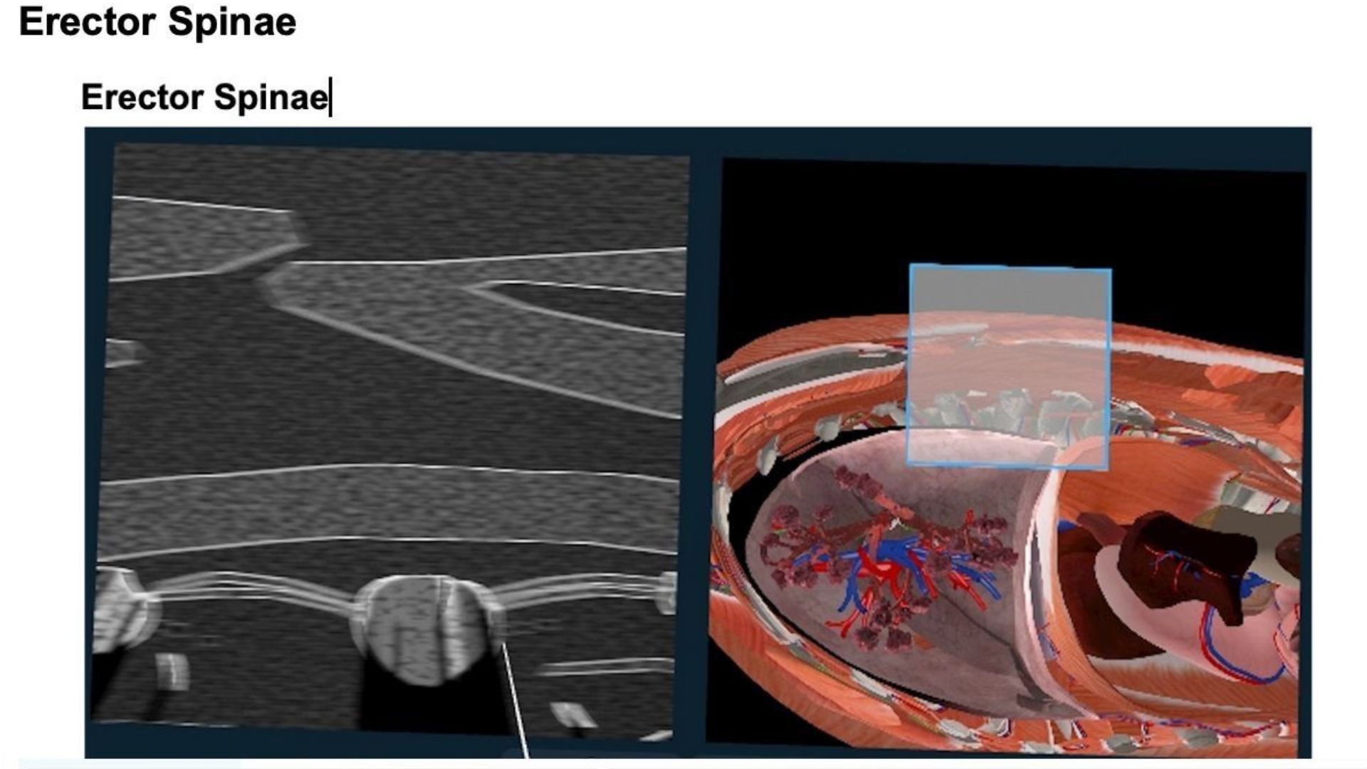
Images from study conducted by the VRiMS Surgical Society research group with Florence Chang and Jason H. leading the project.
Diagnostic Imaging
XR Medical Imaging is transforming diagnostic skills by offering enhanced clarity for interpreting CTs and MRIs. The 3D Organon XR Medical Imaging Module is designed to improve pre-surgical planning and anesthesia management by providing highly detailed, precise visualizations. As a result, healthcare professionals are better equipped to make informed decisions, ultimately improving patient outcomes.
Advanced Tools for Accurate Planning
Several advanced features enhance the accuracy of pre-surgical planning. These include the Pin Tool, which allows for precise annotation of key structures, the Measurement Tool for calculating distances between anatomical landmarks, and the Paint Tool to highlight muscles, bones, and nerves. Together, these tools offer a more focused and accurate approach to visualizing and planning surgical procedures, giving clinicians the ability to identify critical structures with ease.
Enhanced Learning Experience
In addition to improving clinical decision-making, these tools contribute to a richer learning experience. With their ability to provide detailed visualizations, they make it easier to interpret complex anatomical structures. This not only aids in accurate diagnostics but also plays a crucial role in enhancing diagnostic skills and training, leading to better patient outcomes.
Simulation Training for Anesthesiologists
VR simulators are innovative tools designed to help anesthesiologists perfect their guided techniques in a safe, risk-free environment. Whether you’re a beginner mastering the fundamentals or an experienced practitioner fine-tuning your expertise, these simulators provide an immersive, interactive platform to explore human anatomy in detail. Additionally, they offer an excellent opportunity to enhance ultrasound imaging techniques, making them a powerful resource for both new learners and seasoned professionals.
Realistic Simulations for Advanced Skill Development
The 3D Organon VR Ultrasound Simulator delivers highly realistic simulations, offering anesthesiologists the ability to practice and refine their skills in a controlled, virtual environment. For example, it simulates critical procedures such as regional anesthesia and nerve blocks with remarkable accuracy. This technology allows practitioners to hone their skills without the need for real patients, making it an invaluable tool for both ongoing education and hands-on training. As a result, it not only boosts confidence but also ensures that professionals are well-prepared for actual clinical settings.
Advancing Anesthesia Education with 3D Organon
Anesthesiologists, anesthesia professionals, and regional anesthesia experts are increasingly adopting 3D Organon to enhance their practice and refine their skills. This platform offers a highly interactive and realistic simulation environment, enabling perioperative anesthesia specialists to deeply engage with nerve pathways, vascular structures, and muscles. By using 3D Organon, professionals can explore anatomical details with precision—an especially valuable tool in pain management.
Enhancing Pain Management through Advanced Visualization
Clear visualization of anatomical structures is essential for effective block placement and the management of chronic pain. With features like the Slice Tool, XR Imaging, and collaborative Medverse integration, anesthesiologists can engage in real-time, remote discussions with peers and mentors. These capabilities allow for the refinement of techniques, the exchange of insights, and the improvement of patient outcomes in both pain management and overall patient care.
A Game-Changer for Anesthesia Professionals
3D Organon is more than just another tool—it’s a comprehensive platform that is revolutionizing how anesthesiologists approach education and patient care. By enhancing learning experiences and improving clinical outcomes, 3D Organon is making significant strides in advancing both anesthesia education and patient care practices.
Academic and Professional Growth
For students pursuing an academic career, immersive technology offers hands-on research opportunities in healthcare and exposure to innovative methodologies in medical education. This prepares them for scientific research and evidence-based practice.
Case Studies and Industry Leaders Advancing Anesthesia and Pain Medicine
Enhancing Regional Anesthesia Education with VR
Dr. Boyne Bellew, an expert in regional anesthesia and ultrasound-guided nerve blocks, utilizes 3D Organon VR technology to significantly improve trainees’ understanding of anatomy and procedural techniques. By integrating immersive VR tools, he provides a dynamic learning experience that goes beyond traditional methods.
Focusing on Sonoanatomy for Effective Training
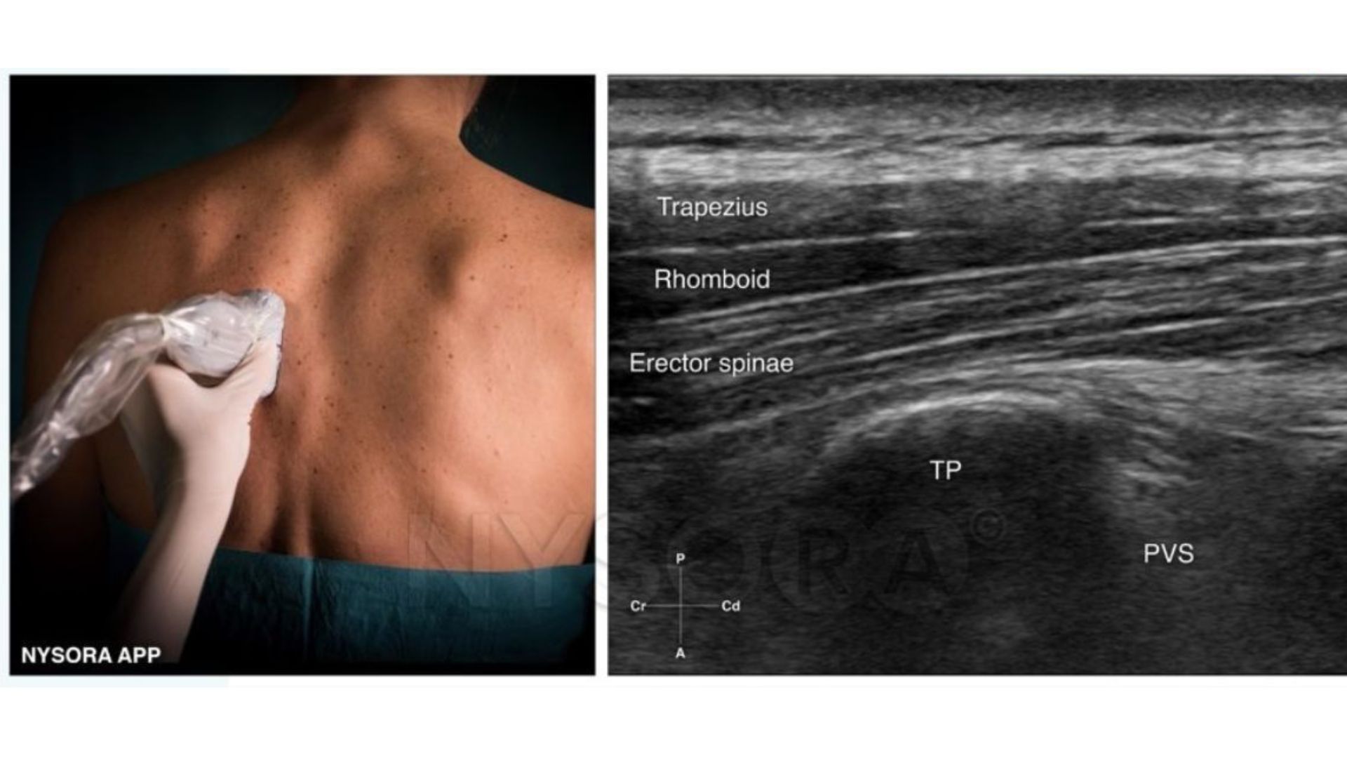
A major focus of Dr. Bellew’s training is sonoanatomy—the study of anatomical structures through ultrasound. In particular, he helps trainees understand nerve pathways, vascular structures, and musculoskeletal relationships, all of which are essential for performing safe and effective regional anesthesia. Through the use of 3D Organon, trainees are able to visualize these structures in detail, which greatly enhances their ability to interpret ultrasound images in real-world settings.
Combining VR with Hands-on Practice
This innovative approach bridges virtual education with hands-on practice. Initially, trainees explore detailed 3D models in the VR environment, and then reinforce their learning using portable ultrasound devices. By combining both methods, Dr. Bellew enhances trainees’ spatial awareness, procedural accuracy, and clinical decision-making, making the training process not only more effective but also more engaging.
Watch the webinar here: 3D Organon Webinar: Teaching Regional Anaesthesia Anatomy/Sonoanatomy using VR
Revolutionizing Patient Care with XR Technology
Dr. Rohan Jotwani, an Interventional Pain Specialist and Anesthesiologist at Weill Cornell, is transforming patient care through the use of 3D Organon’s extended reality (XR) technology. By integrating XR into his practice, Dr. Jotwani enhances collaboration, streamlines procedural planning, and significantly improves patient safety.
Case Study: Enhancing Collaboration Across Distances
In one notable case, Dr. Jotwani and Dr. John Rubin used the 3D Organon Medverse to plan a complex pain management procedure, despite being in different locations. The patient, an elderly individual with intractable pain, required optimal placement for a peripheral nerve stimulator. Thanks to XR technology, both doctors were able to explore the patient’s unique anatomy in greater detail, refining their approach with precision, which traditional imaging methods could not match.
Pre-Surgical Planning: Simulating Procedures for Better Outcomes
For another high-risk case involving a spinal mass, 3D Organon allowed the medical team to simulate the procedure in advance. This simulation helped to optimize needle trajectories and minimize complications. In addition, the risk-free virtual practice environment ensured a safer, more effective intervention, improving patient outcomes.
Watch the Webinar: 3D Organon webinar: XR Opportunities in Pain Medicine – Lessons Learned in Training and Patient Care
Research and Further Resources
Artificial Intelligence-Assisted Virtual Reality Training
- Artificial Intelligence-Assisted Virtual Reality Training Module for Gasserian Ganglion Block: A publication in Interventional Pain Medicine by Oranicha (Natty) Jumreornvong, MD, David Chang, Gonzalo Povea Galdo, Jason Yong, and Rohan Jotwani, MD, MBA. This module enables learners to engage in a 360-degree immersive environment, manipulating holographic anatomy models and simulating fluoroscopic guidance for performing the Gasserian ganglion block.
- Virtual reality training and modeling to aid in pre-procedural practice for thoracic nerve root block in the setting of a schwannoma: A case report published in Interventional Pain Medicine exploring the use of 3D Organon XR for pre-procedural planning for a spine injection near a tumor.
- Artificial intelligence assisted virtual reality training module for Gasserian ganglion block: A proof-of-concept module demonstrating the integration of AI-driven 3D Organon tools with VR to teach advanced procedures, such as the Gasserian ganglion block, a key technique in managing trigeminal neuralgia and severe facial pain.
- Utilisation of extended reality for preprocedural planning and education in anaesthesiology: a practical guide for spatial computing Dr. Rohan Jotwani et al. from the Department of Anesthesiology at NewYork-Presbyterian/Weill Cornell Medicine, explores the transformative potential of extended reality (XR) in healthcare.
- Journal of Medical Extended Reality A leading open-access, peer-reviewed journal dedicated to exploring the role of XR in healthcare.
Newsroom
3D Organon to Participate in the Annual HERDSA Conference 2025
3D Organon to Participate in the Annual HERDSA Conference 2025 3D Organon is excited to announce its participation in the Annual Higher Education...
3D Organon celebrates 10 years of innovation!
3D Organon 10-Year Anniversary July, 2025 - This month, 3D Organon marks a major milestone: 10 years of redefining medical education through...
The Future of Immersive Healthcare Education ft. Dr Panagiota Kordali I Vodafone Business Interview
The Future of Immersive Healthcare Education: Dr. Kordali Shares Insights in Vodafone Business Interview Vodafone Business, in collaboration with...
3D Organon License Management System
Introducing the New 3D Organon License Management System The New Release of 3D Organon introduces a robust set of licensing tools crafted...
3D Organon to be Showcased at AACA 2025!
3D Organon to be Showcased at AACA 2025! We’re proud to announce that the groundbreaking 3D Organon platform will be featured at the 42nd Annual...
3D Organon Featured in Anatomical Sciences Education Journal: Measuring Student Acceptance of VR for Anatomy Learning
3D Organon Featured in Anatomical Sciences Education Journal: Measuring Student Acceptance of VR for Anatomy Learning Anatomy forms the...
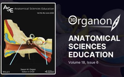
3D Organon Featured on the Cover of Anatomical Sciences Education (ASE)
3D Organon Featured on the Cover of Anatomical Sciences Education (ASE) A 3D Organon model is featured on the cover of the latest issue of...
3D Organon Launches Major Update Featuring AI Study Assistant, Vision Trainer and More
The New 3D Organon Release: Smarter, Faster and More Powerful 3D Organon has officially launched a major update to its leading anatomy learning...
3D Organon at HAPS 2025
Showcasing the Cutting-Edge XR Medical Platform at HAPS 2025 Annual Conference 3D Organon is thrilled to announce its participation in the 39th...
Subscribe to get the latest news!
We promise we don’t send spam
US Office
One World Trade Center, New York City, NY 10007, US
Europe Office
Vas. Sofias, Marousi, Athens, 15124, Greece
Asia | Oceania Office
Township Drive, Burleigh Heads, Gold Coast, QLD 4220, Australia


