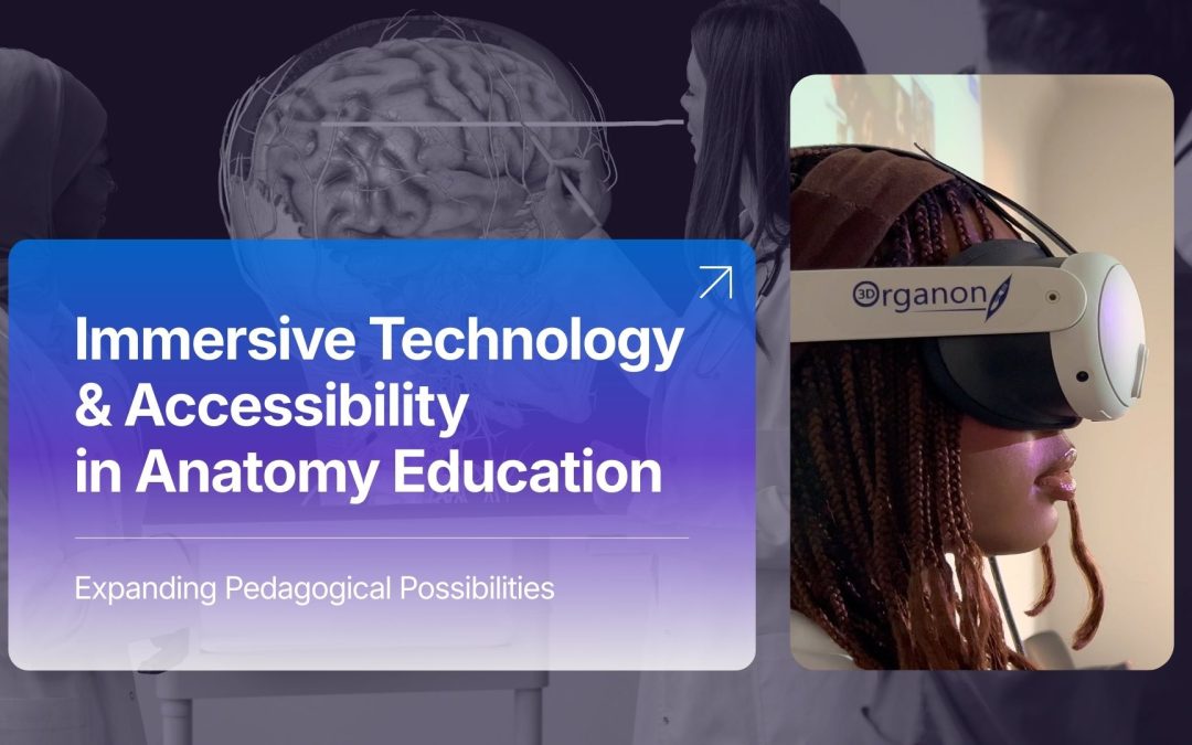
Immersive Technology and Accessibility in Anatomy Education: Expanding Pedagogical Possibilities in Medical Training
“New tech is hard,” remarks Alexi Zukas in a presentation addressing immersive technologies in health sciences education.[1] The statement captures a persistent tension within academic medicine: innovation often meets hesitation not because of its limitations, but because of differences in perception. Attitudes toward extended reality (XR) technologies frequently vary according to generational cohort, educational background and professional culture. While many senior educators remain committed to established instructional models, newer generations, such as Millennials and Gen Z learners, approach immersive technologies with familiarity and curiosity. Generation Alpha, in particular, is developing within an environment in which XR and artificial intelligence (AI) are increasingly normalized components of daily life.
This generational shift underscores a broader pedagogical question: what role should immersive technologies play in anatomy education? Importantly, the discussion should not center on replacement, but rather on accessibility and pedagogical expansion.
The Enduring Role of Traditional Anatomy Instruction
Cadaveric dissection has long been regarded as foundational to medical education. It provides tactile engagement, exposure to anatomical variation, and a profound understanding of human morphology that cannot be entirely replicated through digital simulation. Clinical rotations and patient interactions further contextualize anatomical knowledge within lived human experience.
These traditional modalities, however, are resource-intensive and spatially bound. Cadaver laboratories require substantial infrastructure, ongoing maintenance, regulated access and physical presence. Time within the laboratory is finite, and availability may be constrained by institutional capacity. Moreover, sensory and environmental conditions, such as chemical preservation agents, can present barriers for some learners. Thus, while pedagogically invaluable, conventional methods do not inherently guarantee accessibility.
XR as a Tool for Expanding Access
Digital anatomy platforms have already broadened access to spatial visualization through desktop applications and touchscreen tables. XR technologies extend this development by enabling immersive, manipulable anatomical exploration unconstrained by physical laboratory space.
Within XR environments, learners can isolate anatomical systems, dynamically scale structures and examine spatial relationships in ways that meaningfully complement cadaveric study. Access to high-fidelity anatomical models is no longer confined to a specific laboratory space or timetable; instead, such models can be explored within classrooms, simulation centres, or remote learning contexts. A growing body of empirical research supports the pedagogical value of virtual reality in both in-person and distance education settings. Johnson-Glenberg (2018), identified two primary affordances of VR in educational contexts: an enhanced sense of presence and embodied engagement through gesture-based manipulation of three-dimensional objects. These affordances are particularly relevant to anatomy education, where spatial reasoning, kinesthetic interaction, and contextual immersion are central to meaningful learning.[2]
Pedagogical Frameworks: Differentiated Instruction and Personalized Learning
The integration of XR into anatomy curricula necessitates alignment with established educational frameworks. Two student-centered approaches are particularly relevant: differentiated instruction and personalized learning.
Differentiated instruction is educator-driven and involves adjusting teaching strategies according to students’ readiness, interests, and learning profiles.[3] By offering a variety of pathways through which students can engage with content, educators enhance the likelihood of improved learning outcomes and broaden access to understanding. Within immersive environments, instructors may guide learners through anatomical regions while modulating complexity and focus according to cohort needs.
Personalized learning, by contrast, shifts greater agency to the student. Learners determine aspects of pacing, exploration pathways, and repetition, often supported by adaptive or AI-assisted systems.[4] XR platforms can facilitate repeated engagement with anatomical structures, allowing students to revisit challenging content independently while maintaining academic rigor. Interface adjustments based on individual needs -such as model scalability, alternative display configurations, hand tracking, or seated and standing modes of exploration- further support diverse learning preferences.
These frameworks are not mutually exclusive. Rather, immersive technologies provide a flexible medium capable of supporting both structured instructional guidance and learner autonomy.
A Multi-Modal, Station-Based Approach in Medical Education
A practical application of XR within medical education may be found in a multi-modal, station-based learning model.[5] Consider the teaching of cardiac regional anatomy within a preclinical curriculum. Students might rotate through multiple instructional environments designed to address identical learning objectives through complementary modalities.
One station may involve cadaveric specimens, preserving tactile and spatial authenticity. Another may utilize large digital anatomy tables to facilitate guided small-group exploration under faculty supervision or AI-supported instruction. A third station may immerse students within an XR environment, enabling full three-dimensional exploration of cardiac chambers, coronary circulation, and conduction pathways at adjustable scales. Assessment may follow in digital or traditional formats, reinforcing content mastery across modalities.
Such an approach does not displace traditional instruction; rather, it integrates immersive technology into a multimodal learning ecosystem. The emphasis remains on anatomical competence while expanding methods of engagement.
Accessibility and Equity Considerations
Accessibility in anatomy education extends beyond physical entry into a laboratory. It encompasses cognitive engagement, opportunities for repetition, geographic reach, and accommodation of diverse learner needs. Immersive technology can reduce spatial limitations, support adjustable interfaces, and enable varied modes of interaction, thereby broadening participation in health sciences education.
When immersive platforms align with recognized accessibility standards, they contribute to more inclusive academic environments. In this context, XR should be understood as a mechanism for reducing structural barriers while maintaining educational rigor.
[1] Immersive Education Webinar by 3D Organon: “HSIT Emerging Tech Labs: Redefining Health Sciences Education through XR”, featuring Alexi Zukas, Emerging Technology Labs Coordinator, University of Pittsburgh: https://youtu.be/8FjpjmziRe8?si=N1PJgMLrlQO3ctGJ
[2] Johnson-Glenberg M. C. (2018). Immersive VR and Education: Embodied Design Principles That Include Gesture and Hand Controls. Frontiers in robotics and AI, 5, 81. https://doi.org/10.3389/frobt.2018.0008
[3] Dixon F., Yssel N., McConnell J., Hardin T. (2014). Differentiated instruction, professional development, and teacher efficacy. Journal for the Education of the Gifted, 37(2), 111–127. https://doi.org/10.1177/0162353214529042
[4] Merino-Campos, C. (2025). The Impact of Artificial Intelligence on Personalized Learning in Higher Education: A Systematic Review. Trends in Higher Education, 4(2), 17. https://doi.org/10.3390/higheredu4020017