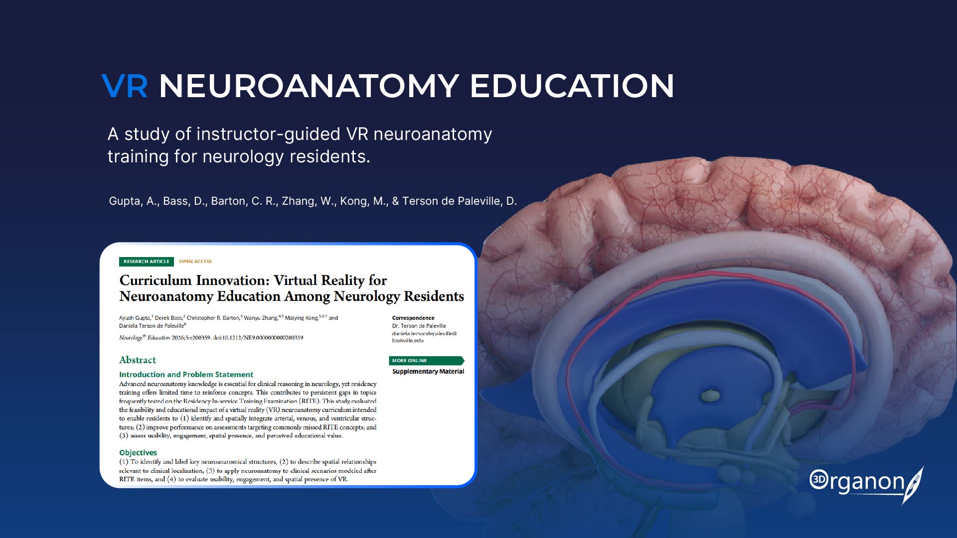
3D Organon XR Supports Neuroanatomy Training for Neurology Residents
A new study published in Neurology® Education is highlighting the potential of virtual reality to support advanced neuroanatomy education in neurology residency training.
The study, “Curriculum Innovation: Virtual Reality for Neuroanatomy Education Among Neurology Residents,” evaluated an instructor-guided virtual reality curriculum developed for neurology and pediatric neurology residents using Organon XR.
Researchers from an academic medical center designed a focused VR learning session around clinically relevant neuroanatomy, including cerebrovascular arterial supply, venous sinuses, ventricular anatomy, and the spatial relationships between these structures.
The findings suggest that integrating 3D Organon XR into a structured, faculty-guided learning experience can support measurable knowledge gains while providing residents with an engaging way to explore complex anatomy.
Addressing a challenge in neurology training
Advanced neuroanatomy plays an important role in neurological clinical reasoning, particularly when interpreting neuroimaging and understanding clinical localization. However, residency programs have limited dedicated time to revisit complex anatomical concepts.
The researchers identified neuroanatomical concepts that residents had found challenging, including material relevant to the Residency In-service Training Examination (RITE), and developed a targeted VR curriculum around these areas.
Rather than relying on two-dimensional representations alone, the curriculum used Organon VR Anatomy to give residents an immersive environment in which they could directly explore anatomical structures and their spatial relationships.
A focused VR session with 3D Organon
Thirty neurology and pediatric neurology residents participated in the study.
During the approximately 40-minute session, residents used Organon VR Anatomy on Meta Quest 3 headsets under faculty guidance. The curriculum focused on intracranial arterial and venous anatomy as well as the ventricular system.
The VR environment was adapted to the educational objectives, allowing learners to focus on the structures and relationships most relevant to the session.
Faculty members guided the experience through questioning, feedback, and clinical contextualization, connecting the anatomical structures residents encountered in VR with clinical scenarios.
Knowledge was assessed at three points: before the session, immediately afterward, and again two weeks later.
Knowledge gains persisted at follow-up
The study found measurable improvement in residents’ assessment performance following the VR intervention.
Mean scores increased from 8.57 at baseline to 11.37 immediately after the session. At the two-week follow-up, the mean score was 11.20, remaining substantially above the baseline result.
The findings suggest that the improvement was not limited to the immediate post-session assessment and that residents retained much of the knowledge gained during the intervention.
The researchers also observed differences according to training level, with more senior residents demonstrating greater improvement, while junior residents entered the session with higher baseline scores.
Residents reported a positive VR experience
Alongside the knowledge assessments, the researchers evaluated residents’ perceptions of the VR experience.
Participants reported high levels of enjoyment, engagement, spatial presence, and usability. The study also found no significant reported cyber fatigue following the session.
The results indicate that immersive anatomy can be incorporated into a relatively short educational session without requiring residents to spend extensive additional time away from their clinical responsibilities.
The role of 3D Organon in the learning experience
For complex neuroanatomy, one of the potential advantages of immersive 3D technology is its ability to make spatial relationships directly explorable.
With 3D Organon VR, learners can interact with anatomical structures and examine their relationships from different perspectives. In the context of this study, this capability was used to support understanding of neurovascular and ventricular anatomy that can be difficult to represent fully through conventional two-dimensional resources.
The researchers’ approach also demonstrates the importance of combining the technology with expert instruction. The VR environment provided the anatomical visualization and interaction, while faculty guided residents through the material and connected it to clinical reasoning.
The study therefore positions VR as a complement to expert teaching, rather than a replacement for it.
From anatomy education to clinical application
The study is notable not only because it evaluates VR, but because it applies immersive anatomy education to graduate medical education.
Much of the research surrounding VR anatomy has focused on undergraduate medical learners. In residency, however, the educational challenge shifts from acquiring foundational anatomical knowledge to applying that knowledge in increasingly complex clinical contexts.
For neurology residents, understanding three-dimensional relationships between arteries, veins, ventricles, and surrounding structures can contribute to the interpretation of neuroimaging and clinical localization.
The use of 3D Organon VR within this curriculum reflects this progression — from learning where structures are to understanding how their spatial relationships relate to clinical scenarios.
Implementation matters
The study also highlights several practical considerations for institutions introducing VR into medical education.
Running multi-headset sessions requires reliable connectivity and appropriate IT support, while institutional firewalls and security systems can create technical challenges. Faculty also need time to prepare and facilitate sessions and to adapt content to different levels of learner experience.
These considerations reinforce the importance of treating VR not simply as a piece of hardware, but as part of a broader educational system.
A growing role for immersive medical education
The findings add to growing research into how immersive technologies can be incorporated into medical education beyond traditional anatomy instruction.
For 3D Organon, the study provides an example of Organon VR Anatomy being used in a real graduate medical education setting to support advanced neuroanatomy training.
While the researchers describe the findings as preliminary and call for further controlled research, the study demonstrates the feasibility of integrating instructor-guided VR into residency education and provides evidence of improved short-term knowledge outcomes.
As medical education continues to explore immersive technologies, applications such as this point toward a role for VR that goes beyond simply visualizing anatomy: helping learners explore complex structures in three dimensions and connect that understanding to clinical reasoning.
Read the full study
Gupta A, Bass D, Barton CR, Zhang W, Kong M, Terson de Paleville D. “Curriculum Innovation: Virtual Reality for Neuroanatomy Education Among Neurology Residents.” Neurology® Education. 2026.
Explore what’s new in 3D Organon XR and discover how immersive anatomy can become an even more powerful part of medical education. Contact us at [email protected] to start your immersive journey.