
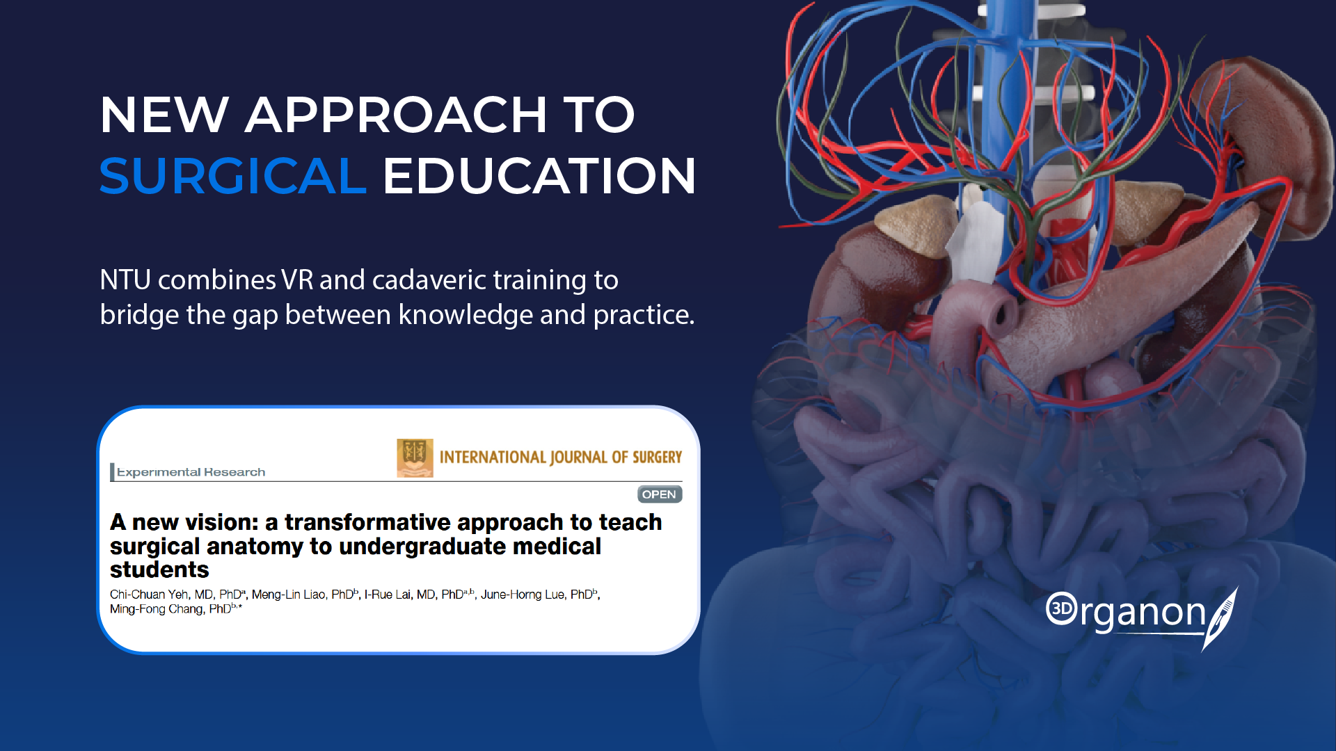
New Approach to Surgical Education: NTU Combines VR and Cadaver Practice to Teach Surgical Anatomy
A new elective course at National Taiwan University combines 3D Organon XR, and hands-on cadaveric training to help medical students bridge the gap between anatomical knowledge and real-world surgical practice.
Medical students spend years studying human anatomy, but recognizing anatomical structures in an actual surgical setting presents an entirely different challenge. This is particularly true in laparoscopic surgery, where surgeons must navigate complex anatomical regions through a restricted, camera-based view.
Researchers at National Taiwan University (NTU) have developed an innovative teaching approach designed to address this challenge. By combining structured anatomy lectures, immersive virtual reality (VR) simulations powered by 3D Organon, and practical training on donated human bodies, the course gives students an opportunity to experience surgical anatomy from multiple perspectives.
The research was led by Chi-Chuan Yeh, alongside Ming-Fong Chang and a multidisciplinary team of surgeons and anatomists at National Taiwan University. Published in the International Journal of Surgery (2026; 112:643–654), the study evaluated the elective course Clinical Abdominal Surgical Anatomy over two academic years, examining how combining VR technology with traditional anatomical teaching and hands-on surgical practice could improve students’ understanding of surgical anatomy.
Bridging the Gap Between Anatomical Knowledge and Surgical Practice
Traditional anatomy education provides students with a strong foundation through textbooks, anatomical illustrations, and cadaveric dissection. However, translating this knowledge into clinical practice can be challenging.
In particular, laparoscopic surgery presents a distinct learning curve. Unlike open surgery, where anatomical structures can be examined directly, laparoscopic procedures rely on a camera that provides a restricted, magnified view of the surgical field.
The NTU research team identified this gap as an important challenge in undergraduate medical education. Students often struggle to translate what they have learned through conventional anatomy teaching and open cadaveric dissection into the perspective encountered during minimally invasive procedures.
The course, titled Clinical Abdominal Surgical Anatomy, was designed to help students make this transition before entering more advanced clinical training.
A Three-Step Learning Experience Powered by VR
Developed collaboratively by surgeons and anatomists, the course integrated three complementary teaching methods to help students understand the relationship between anatomical structures and their appearance during surgery.
- Revisiting the Anatomical Foundations
Each session began with a focused review of relevant anatomy, connecting previously acquired knowledge from gross anatomy courses with the specific anatomical regions involved in the upcoming surgical procedures.
- Experiencing the Laparoscopic Perspective Through VR
Students then explored first-person VR simulations designed to recreate the visual perspective of laparoscopic surgery.
Using HTC VIVE Pro 2 headsets and 3D Organon XR software, instructors demonstrated how anatomical structures appear during minimally invasive procedures. The platform’s layer-penetrating functionality allowed educators to reveal underlying anatomical structures and illustrate the relationship between different tissue layers, helping students understand what they would encounter through a laparoscope.
This immersive experience provided an additional perspective on anatomy, connecting the three-dimensional relationships of anatomical structures with their appearance during surgical procedures.
- Learning Through Real Surgical Videos
The final lecture component involved unedited surgical videos narrated by experienced surgeons. Students followed the procedures, identified important anatomical structures, and marked them on an interactive touch display.
By combining anatomical explanations with actual surgical footage, the course helped students connect theoretical knowledge and VR simulations with the complexity of real clinical procedures.
Taking Learning Beyond the Classroom
The educational experience continued in the anatomy laboratory, where students participated in four-hour practical sessions using formalin-fixed donated bodies, referred to by the program as silent mentors.
Working in small groups under the guidance of surgical residents or attending surgeons, students rotated through different operating room roles, including operator, assistant, scrub nurse, and circulating nurse.
The practical sessions covered three surgical procedures: Inguinal hernia repair, Cholecystectomy with partial liver resection, and Gastrectomy.
This approach allowed students to explore surgical anatomy while also gaining a broader understanding of teamwork, procedural workflows, and the responsibilities of different operating room professionals.
Significant Improvements in Student Performance
The researchers evaluated the course over two academic years, involving 36 medical students in total.
The results showed substantial improvements in anatomical knowledge following the lectures. Across all three surgical procedures, average test scores increased from approximately 6.2–6.4 out of 10 before instruction to 9.8–9.9 after instruction.
Students also responded positively to the overall learning experience. In a 20-item questionnaire, most aspects of the course received ratings between 4.3 and 4.9 on a five-point scale.
The anatomy review received the highest average rating (4.92), followed by the surgeon-narrated surgical videos (4.83). The VR component also received a favorable rating of 4.31.
Beyond knowledge acquisition, students reported increased confidence in learning other surgical procedures and a greater understanding of the operating room environment. The researchers also noted that 97–100% of students endorsed the VR simulations in the procedures for which feedback was collected.
These findings suggest that combining conventional anatomy teaching with immersive technology and hands-on experience can create a more comprehensive learning environment for future medical professionals.
The Future of Immersive Surgical Anatomy Education
The NTU study highlights the value of integrating different educational approaches to address the challenges of modern surgical training.
Rather than replacing traditional anatomy education, VR provides an additional layer of understanding, allowing students to explore anatomical relationships from perspectives that are difficult to reproduce through textbooks or conventional dissection alone.
The research team plans to continue developing the course, with potential improvements including independent VR learning sessions, the use of fresh cadavers for laparoscopic practice, and larger multicenter studies to evaluate the approach further.
For medical education, the implications extend beyond a single elective course. As minimally invasive procedures become increasingly important in modern healthcare, helping students understand anatomy from the perspective of the surgeon may become an essential part of preparing them for clinical practice.
Read the full study
Yeh, C. C., Liao, M. L., Lai, I. R., Lue, J. H., & Chang, M. F. (2026). A new vision: a transformative approach to teach surgical anatomy to undergraduate medical students. International journal of surgery (London, England), 112(1), 643–654.
Explore what’s new in 3D Organon XR and discover how immersive anatomy can become an even more powerful part of medical education. Contact us at [email protected] to start your immersive journey.